Human consciousness and psychology are, in essence, functions of the human brain. Whenever we investigate a question, we are likely to encounter the field of brain science, so some basic knowledge is important. The following is a summary of key concepts. Reading it through and returning to it from time to time can offer valuable insights for learning.
I. What Is Brain Science?
Brain science is essentially neuroscience, which studies the human nervous system. The nervous system is one of the body’s systems: it controls muscle activity, coordinates tissues and organs, establishes and receives information from outside the body, and coordinates it. We rely on it to sense external information, process that information, and control the body’s response.
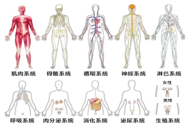
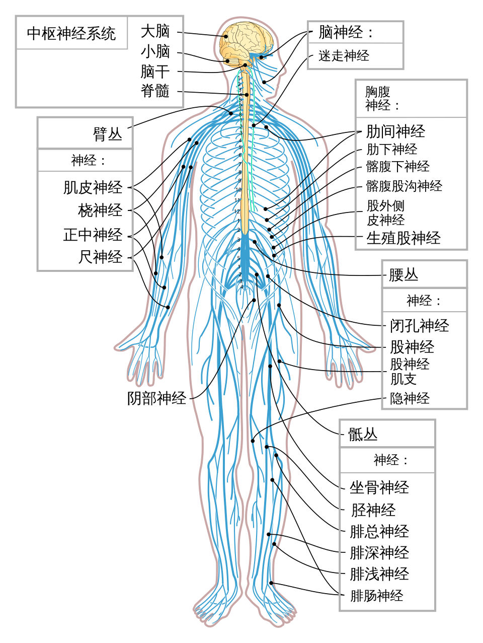
At the broadest level, the nervous system is divided into the central and peripheral nervous systems. The former includes the brain and spinal cord; the latter includes the nerves connecting the central nervous system with different parts of the body, such as the cranial and spinal nerves.
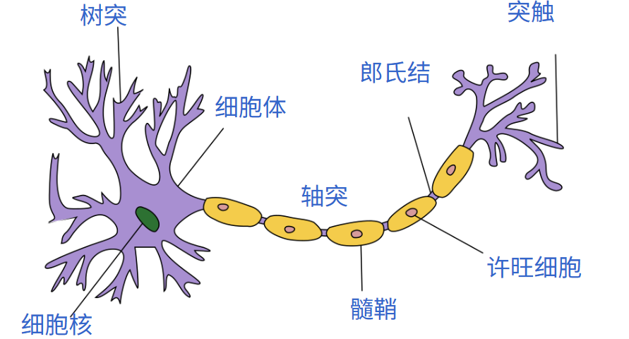
At the cellular level, the nervous system consists of neurons, or nerve cells, and glial cells. Neurons transmit signals and carry out the nervous system’s functions. Glial cells come in many varieties and mainly support, nourish, and protect neurons. The human brain is estimated to contain about 85–120 billion neurons, with a similar or slightly greater number of glial cells.
Problems in certain nerves can cause numbness in the legs and feet. Problems in the middle of the spinal cord can cause paralysis on one side of the body. Problems in the brain may produce symptoms such as dementia. When the brain loses all function, a person’s consciousness disappears. Medically, this is brain death, and we can essentially say that the person has died too.
On the positive side, when we describe someone as having a high IQ, high emotional intelligence, a strong memory, or broad knowledge and experience, we are really saying that some function of their brain is particularly strong. By consciously caring for and exercising our brains, we too can become more intelligent, enjoy richer mental lives, and live more happily.
II. The Structure of the Brain
The brain’s structure is complex, and research is not yet comprehensive. The following divides the brain according to its anatomical structures:

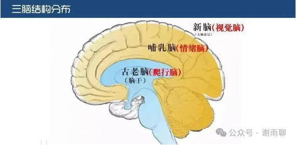
The triune brain is another important theoretical model of brain structure. It is not accurate, but it is highly practical. It divides the human brain into the reptilian brain, the mammalian brain—the limbic system—and the human brain—the neocortex. The outer layers are more advanced, and the inner layers more basic. These can also be understood as the instinctive, emotional, and rational brains. Their divisions and functions are shown below:
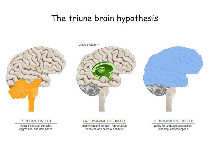

III. Functions of Important Brain Regions
The following is an incomplete list of the functions of important brain regions, intended as a quick reference.
1. Brainstem
-
Location: At the base of the brain, connecting it to the spinal cord; the “life center” of the entire nervous system.
-
Main functions: Controls life-sustaining functions such as heart rate, breathing, blood pressure, body temperature, and digestion, and regulates sleepiness and the alarm system.
-
Implications: Most brainstem functions operate on “autopilot” and cannot be directly changed through willpower. Breathing is the one “control switch.” The frontal motor area and hypothalamus both help regulate breathing. Abdominal breathing can stimulate the release of endorphins and serotonin and is the most direct physical method for quickly calming the body’s stress response.
2. Cerebellum
-
Location: Behind the brainstem and below the cerebral hemispheres.
-
Main functions: Maintains balance, muscle coordination, precision of movement, and the formation of conditioned reflexes.
-
Implications: Muscle memory and skilled performance depend on the cerebellum. When repeated practice makes a skill—such as playing ball or cycling—“automatic,” the cerebellum and basal ganglia have taken over the task, freeing the prefrontal cortex’s processing resources.
3. Amygdala
-
Location: Within the limbic system, closely adjacent to the hypothalamus.
-
Main functions: Processes intense emotions such as fear, anger, and excitement, triggering the “fight or flight” response. It is a meeting point of the human, mammalian, and reptilian brains, and a “transfer station” between reason and emotion.
-
Implications: When confronted with intense fear or threat, the amygdala can instantly “hijack” the brain, depriving the entire frontal lobe of its ability to think—“fear makes intelligent people stupid.” The amygdala cannot be trained simply by reading; it requires firsthand experience and actual practice. Successfully confronting difficulties provides lifelong resources for reshaping its response patterns.
4. Hypothalamus
-
Location: Below the thalamus, closely adjacent to the amygdala.
-
Main functions: Regulates body temperature, thirst, hunger, blood pressure, heart rate, the sleep–wake clock, nurturing instincts, and sexual arousal; it also participates in the brain’s reward system.
-
Implications: The hypothalamus governs basic human drives and internal homeostasis. Deliberately practicing healthy routines and habits can effectively “train” its responses, making the body’s physiological rhythms more stable.
5. Hippocampus
-
Location: Within the medial temporal lobe, in a core region of the limbic system.
-
Main functions: Converts working memory into long-term memory, especially episodic memory, and participates in spatial navigation.
-
Implications: The hippocampus is a memory “transfer station,” not a “storage vault.” Emotional fluctuations and deep sleep are crucial to its consolidation of memories. Novel environments and positive emotions can significantly enhance hippocampal neuroplasticity.
6. Cingulate Gyrus
-
Location: On the medial surface of the cerebral hemispheres, wrapping around the corpus callosum; an important part of the limbic system.
-
Main functions: Participates in emotional processing, the expression of anxiety, pain perception, self-regulation, and negative imagination.
-
Implications: The cingulate gyrus is usually more active in women than in men, making women more sensitive to emotions and potential risks and more prone to anxiety, but also giving them greater potential for emotional regulation. Recognizing “negative imagination” as simply a physiological response of the cingulate gyrus can help us practice mindfulness and self-control more effectively.
7. Frontal Lobe
-
Location: In the front part of the brain; the largest cerebral lobe.
-
Main functions:
Prefrontal cortex: planning, analysis, judgment, concentration, self-control, social interaction, and abstract thought. It is the last brain region to mature, usually reaching full maturity around age 25.
Broca’s area: responsible for language production and contains mirror neurons.
Premotor area and primary motor cortex: control bodily movement and connect to the brainstem.
- Implications: Deliberate practice essentially means using the human brain—the prefrontal cortex—to train the mammalian and reptilian brains. Because modern people process huge amounts of information every day, the prefrontal cortex tires easily, reducing self-control. Be more understanding toward young people, whose prefrontal cortex is not fully developed. At the same time, make good use of mirror neurons: learning through imitation and personal example is far more efficient than purely theoretical preaching.
8. Parietal Lobe
-
Location: At the top of the brain, behind the frontal lobe.
-
Main functions: Integrates bodily sensory information such as touch; supports spatial imagination, abstraction, mathematical calculation, and movement coordination. It is associated with overall coordination and sudden insight.
-
Implications: The parietal lobe gives humans strong spatial perception and abstract thinking. When we construct a complex model in our minds or experience a “flash” of insight, the parietal lobe is often working in the background to integrate multiple kinds of information at a broad level.
9. Temporal Lobe
-
Location: On both sides of the brain, above the ears.
-
Main functions:
Wernicke’s area: located in the superior temporal gyrus and responsible for language comprehension.
Middle and inferior temporal gyri: responsible for auditory processing, understanding meaning, facial recognition—memory for faces—and the formation of long-term memories.
- Implications: The human brain’s capacity to process visual information far exceeds its capacity for auditory information. Simply “listening to a lecture” has limited efficiency. “Drawing while explaining,” as in the Feynman learning technique, transforms auditory information into visual images and can more efficiently activate the temporal lobe and memory across the brain.
10. Occipital Lobe
-
Location: In the lower rear part of the brain, at the back of the head.
-
Main functions: Specializes in higher-level processing and analysis of visual information and contains the primary visual cortex.
-
Implications: “A picture is worth a thousand words” has a solid neuroscientific foundation. The human brain is extremely powerful at processing visual information. Using more charts, mind maps, and concrete images when learning, communicating, or sharing knowledge can greatly reduce the effort required for understanding and improve memory.
IV. Brain Development
The brain does not stop changing in adulthood. Its development proceeds in stages.
Different abilities “peak” at different ages. Research from the Massachusetts Institute of Technology (MIT) and Harvard University shows that brain functions do not all decline after age 20; instead, different functions peak at different stages of life:
Ages 18–19: information-processing speed and reaction ability peak.
Around age 25: short-term memory—working memory—peaks.
Around age 30: the ability to recognize and remember unfamiliar faces peaks.
Ages 40–50: understanding others’ emotions—empathy—and social insight peak.
Ages 50–60: vocabulary, the integrated application of knowledge, and the ability to solve complex real-world problems peak.
It is particularly important to note that brain development progresses from back to front. The prefrontal cortex, responsible for emotional control, long-term planning, and risk assessment, is the last region to mature, usually around age 25. So statements such as “young people are impulsive” and “young people’s brains are not fully developed” have a scientific basis.
The brain also possesses lifelong neuroplasticity. Even in adulthood, we can still improve it. Whenever we learn a new skill or change a habit of thought, new connections—synapses—form between neurons. Frequently used neural pathways become “myelinated,” like turning a country lane into a highway, making responses faster and faster. Connections left unused for a long time are “pruned” by the brain. Age is therefore never an excuse to stop learning. With continuous small stimuli and learning across fields, the brain can still rebuild its networks and remain sharp into middle and old age.
V. Small Ways to Improve Brain Function
As discussed above, the brain possesses lifelong neuroplasticity. On the negative side, drinking permanently changes the brain, drug use permanently changes it, and depression permanently changes it. All of these statements are true. But we can also use this principle in the other direction, taking measures to change the brain for the better. Here are a few small methods:
1. Do 150 Minutes of Moderate Aerobic Exercise Each Week
Promotes the release of brain-derived neurotrophic factor (BDNF), directly stimulating the hippocampus to produce new neurons.
2. Get Enough Deep Sleep
Activates the brain’s “lymphatic clearance system,” removes waste proteins produced by daytime metabolism, and consolidates long-term memory in the hippocampus.
3. Eat an Anti-inflammatory Diet: Omega-3, Antioxidants, and Fewer High-sugar and Processed Foods
Reduces chronic inflammation in the brain and protects the fluidity of neuronal cell membranes.
4. Learn New Skills Across Different Fields
Breaks established patterns of thinking and creates entirely new synaptic connections between neurons.
5. Think Actively and Explain What You Know, as in the Feynman Technique
Forces the prefrontal cortex to integrate information deeply, turning it into durable long-term neural circuits.
6. Practice Mindfulness and Concentration
Increases the thickness of the prefrontal cortex, reduces excessive amygdala activity, and strengthens emotional self-control.
7. Change Everyday Habits to Provide Small Stimuli
Brush your teeth with your non-dominant hand or take a different route home, forcing the brain to switch from “autopilot” to “active awareness.”
Comments
No account needed. A nickname is required; email is optional. Successful submissions appear publicly without manual pre-moderation.
Comments and nicknames are public. Optional email addresses are not displayed. The selected Waline backend and its hosting provider process comments, nicknames, optional email, IP and browser information. Comments show IP region, without the full IP address or browser and operating system labels. Hosting: Vercel; database: Neon.